


· Consistent and reproducible results
· No heavy & light antibody chains
· Extraordinary binding, even under harsh conditions
· High affinity
· Short Incubation (5-30 min)
Description:
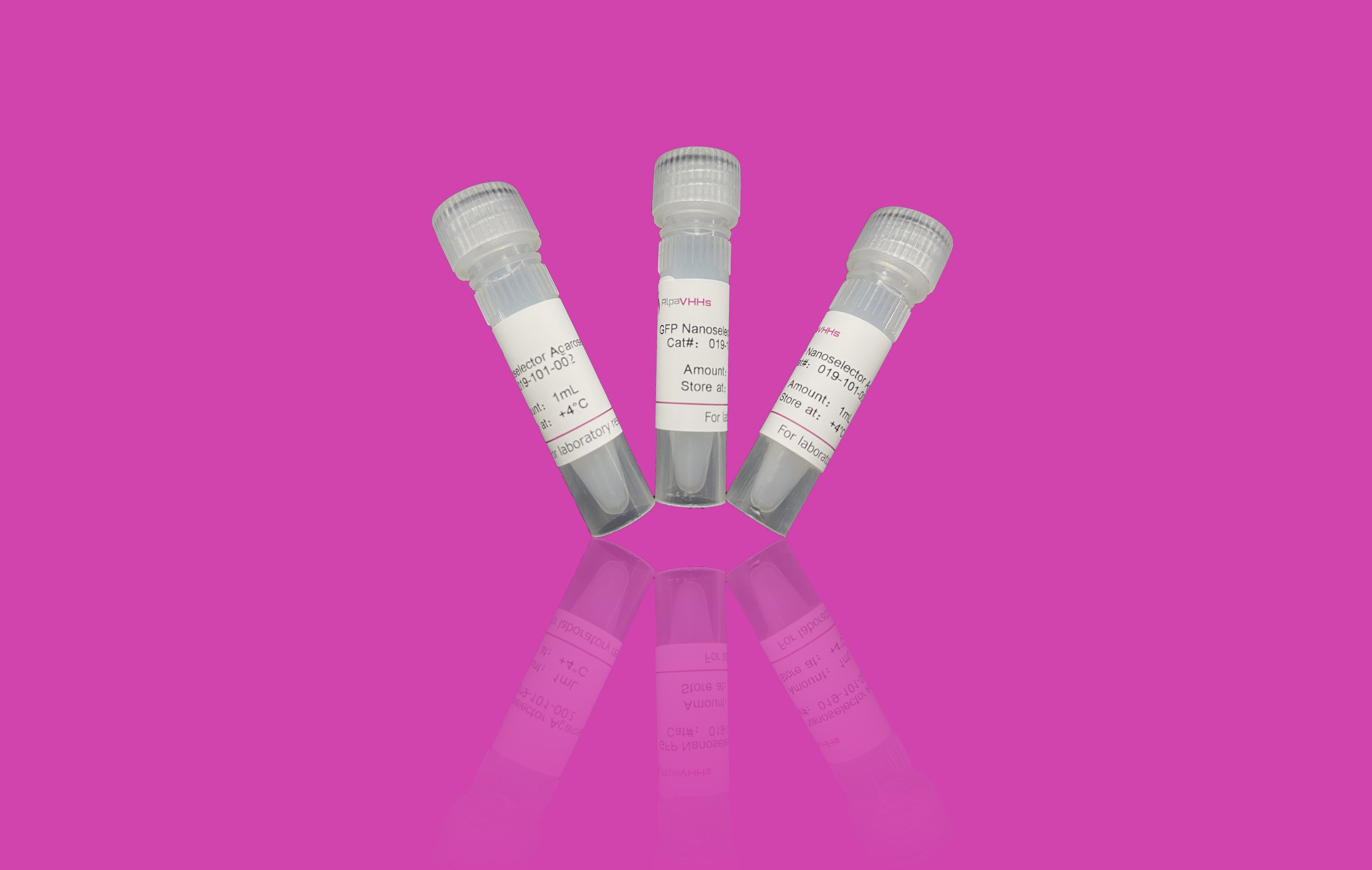
GFP Nanoselector Agarose have been specifically designed to bind GFP-fusion proteins. GFP Nanoselector Agarose is based on small high-affinity recombinant alpaca antibody fragments covalently coupled to the surface of Agarose. GFP Nanoselector Agarose is an ideal tool to isolate or purify GFP-fusion proteins fast and efficiently.
Ligand: Anti-GFP single domain antibody fragment (VHH, Nanobody)
Bead size: ~ 40µm
Reactivity: Recognizes GFP, mEGFP, superfolder GFP and most common CFP and YFP variants. Does not cross-react with mCherry, mRFP, dsRed, mTagBFP, mTagRFP or their most common derivatives.
Binding capacity: High binding capacity, 10 µL slurry bind about 20 µg of recombinant GFP.
Storage: Shipped at ambient temperature. Upon receipt store at 4°C. Stable for 1 year. Do not freeze.
Storage Buffer: 50 % slurry in PBS containing 20 % Ethanol
Background:
Green fluorescent proteins (GFPs) and the variants thereof are widely used to study protein localization and dynamics. For biochemical analysis including mass spectrometry and enzyme activity measurements these GFP-fusion proteins and their interacting factors need to be isolated fast and efficiently by immunoprecipitation using the GFP Nanoselector Agarose. Due to the single-chain nature of sdAbs and their stable and covalent attachment, no leakage of light and heavy chains is observed during elution with SDS sample buffer. GFP Nanoselector is compatible not only with physiological buffers but also with high stringency buffers which provides great freedom to adjust the binding and washing conditions to the experimental needs.
Immunoprecipitation (IP)/Co-IP
Mass spectrometry (MS)
Enzyme activity measurements
Immunoprecipitation protocol
Mammalian cell lysis
Note: Harvesting of cells and cell lysis should be performed with ice-cold buffers. We strongly recommend to add protease inhibitors to the Lysis buffer to prevent degradation of your target protein and its binding partners.
For one immunoprecipitation reaction, we recommend using ~106- 107 cells.
1. Choice of lysis buffer:
* For cytoplasmic proteins, resuspend the cell pellet in 200 µL ice-cold Lysis buffer by pipetting up and down. Supplement Lysis buffer with protease inhibitor cocktail and 1 mM PMSF (not included).
* For nuclear/chromatin proteins, resuspend cell pellet in 200 µL ice-cold RIPA buffer supplemented with DNaseI (f.c. 75-150 Kunitz U/mL), MgCl2 (f.c. 2.5 mM), protease inhibitor cocktail and PMSF(f.c. 1 mM)(not included)
2. Place the tube on ice for 30 min and extensively pipette the suspension every 10 min.
3. Centrifuge cell lysate at 17,000x g for 10 min at +4°C. Transfer cleared lysate (supernatant) to a pre cooled tube and add 300 µL Dilution buffer supplemented with 1 mM PMSF and protease inhibitor cocktail (not included). If required, save 50 µL of diluted lysate for further analysis (input fraction).
Bead equilibration
1. Resuspend the beads by gently pipetting up and down or by inverting the tube. Do not vortex the beads!
2. Transfer 25 µL of bead slurry into a 1.5 mL reaction tube.
3. Add 500 µL ice-cold Dilution buffer.
4. Sediment the beads by centrifugation at 2,500x g for 5 min at +4°C.
5. Discard the supernatant.
Protein binding
1. Add diluted lysate to the equilibrated beads.
2. Rotate end-over-end for 1 hour at +4°C.
Washing
1. Sediment the beads by centrifugation at 2,500x g for 5 min at +4°C.
2. If required, save 50 µL of supernatant for further analysis(flow-through/non-bound fraction).
3. Discard remaining supernatant.
4. Resuspend beads in 500 µL Wash buffer.
5. Sediment the beads by centrifugation at 2,500x g for 5 min at +4°C. Discard the remaining supernatant.
6. Repeat this step at least twice.
7. During the last washing step, transfer the beads to a new tube.
Optional: To increase stringency of the Wash buffer, test various salt concentrations e.g. 150 mM - 500 mM,and/or add a non-ionic detergent e.g. Triton™ X-100.
Elution with 2x SDS-sample buffer
1. Remove the remaining supernatant.
2. Resuspend beads in 80 µL 2x SDS-sample buffer.
3. Boil beads for 5 min at +95°C to dissociate immunocomplexes from beads.
4. Sediment the beads by centrifugation at 2,500x g for 2 min at +4°C.
5. Analyze the supernatant in SDS-PAGE.
Elution with Glycine-elution buffer
1. Remove the remaining supernatant.
2. Add 50–100 µL Glycine-elution buffer and constantly pipette up and down for 30 - 60 sec at +4°C.
3. Sediment the beads by centrifugation at 2,500x g for 5 min at +4°C.
4. Transfer the supernatant to a new tube.
5. Immediately neutralize the eluate fraction with Neutralization buffer.
6. Repeat this step at least once to increase elution efficiency .
